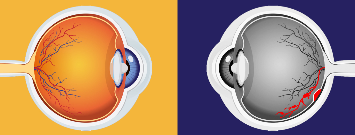
What is Macular Edema?
When blood vessels in the macula become damaged and leak, it causes swelling. This is called macular edema. The retina is a light-sensing tissue located at the back of the eye. The central part of the retina is called the macula. The macula gives us the detailed, colorful vision needed for reading, driving, and distinguishing faces. Macular edema causes washed out colors and blurred vision. Without treatment it may lead to permanent vision loss.1,2
Causes of macular edema
Many health conditions may cause the leakage that leads to swelling in the retina, or macular edema. These are:
- Diabetes
- Age-related macular degeneration (AMD)
- Macular holes or puckers
- Retinal vein occlusion (RVO)
- Hereditary disorders (passed from parent to child), such as retinoschisis or retinitis pigmentosa
- Inflammatory eye diseases
- Certain medicines
- Eye tumors
- Eye surgery
- Eye injuries1,2
Symptoms of macular edema
The most common symptom of macular edema is blurry or wavy vision in the center of your vision. Colors may also seem washed out, faded or different. It is painless. If only one eye is affected, you may not notice any blurriness until the condition is advanced.1
Diagnosing macular edema
An optometrist or ophthalmologist can diagnose macular edema during a thorough eye exam. The tests they use will help determine the location and severity of the swelling, and include1,2:
Visual acuity test
For this test, your doctor has you look at a chart with rows of letters, with and without your glasses or contacts, to identify how well you see. You will be asked to read out loud the smallest line of letters you can see, using both eyes, and one eye at a time.
Dilated eye exam
Drops are placed in your eyes to widen, or dilate your pupils. This allows your doctor to see the retina (back of the eye) more easily and judge the retina’s health. This exam may reveal blood vessel leaks, holes or cysts.
Fluorescein angiogram
If your doctor suspects macular edema, they may want to perform this additional test. A yellow dye (called fluorescein) is injected into a vein, usually in your arm. The doctor then uses a special camera to take photos of the dye as it travels through the blood vessels in the back of your eye. This test allows your doctor to identify the amount of damage to the macula.
Optical coherence tomography (OCT)
Similar to an ultrasound, this test uses a special light and camera to take detailed cross-sectional pictures of your retina and macula. OCT detects the thickness of the retina, which can indicate the amount of swelling.
Amsler grid
You can use an Amsler grid at home to detect even small changes in your vision. You should discuss any vision changes with your doctor as soon as possible.
Treatments for macular edema
The type of treatment you receive will depend greatly on what is causing the swelling. These treatments may include1,2:
Anti-VEGF drugs
This is the most common treatment for macular edema. Anti-VEGF drugs are injected into the eye at regular intervals. These medications help reduce the growth of abnormal blood vessels underneath the retina and slows any leaks from those vessels, so your eye has time to resorb any existing fluid. Anti-VEGF drugs are also prescribed for wet AMD.
Steroids
If your macular edema is caused by inflammation, your doctor may prescribe steroids delivered as eye drops, pills or injections.
NSAID eye drops
For macular edema that occurs after surgery for cataracts, glaucoma or retinal disease, your doctor may prescribe non-steroidal anti-inflammatory eyedrops for a few months.
Vitrectomy
During a vitrectomy, the doctor removes the vitreous gel (the substance that fills the eye and gives it a round shape) and replaces it with a bubble of air and gas. This bubble pushes against the edges of the macular hole, allowing it to heal.
Laser surgery
Until recently, focal laser photocoagulation was a common treatment for macular edema. However, recent research has shown that anti-VEGF injections are more effective.
Diabetes control
One of the common complications of diabetes is a condition called diabetic retinopathy. Diabetic retinopathy damages the small blood vessels of the retina which in turn causes fluid to leak into the retina which leads to swelling of the macula. That’s why, if you have diabetic macular edema, your doctor will likely recommend you try to control your blood sugar and blood pressure more closely to help reduce further blood vessel damage.

Join the conversation