In order to understand macular degeneration, you first need to get a basic idea of the general anatomy of the healthy eye. The eye is the organ that helps us see, and though it may look like one whole, there are many different parts to the eye. Knowing which parts are responsible for what can help you better understand just how sight operates, and what happens when something goes awry, like in macular degeneration.
How do we see?
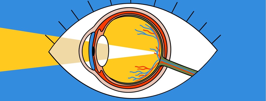
Sight is intricately linked to light. Light enters the eye through the cornea, then travels through the lens.1 These together help to focus the light onto the retina, which is located in the back of the eye. The retina then uses special light-sensing cells, called photoreceptors, to send signals to the brain via the optic nerve.1 The brain then interprets these signals, giving you an image.
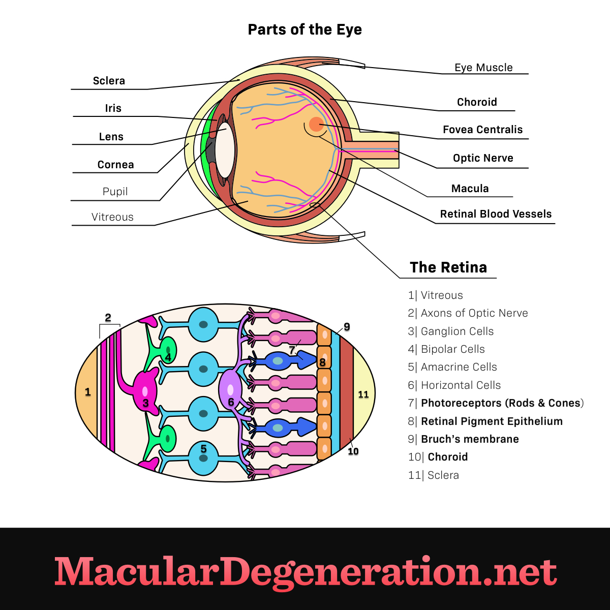
Figure 1. The anatomy of the eye
Parts of the outer eye
- Sclera: The sclera is the protective white outer layer of the eye.1
- Iris: The iris is the colored part of the eye, with the pupil in the center of it. The pupil dilates and contracts depending upon the amount of light that is present.1
- Lens: The lens further focuses light onto the retina.1
- Cornea: The cornea can be compared to the windshield of a car. It is located at the front of the eye and is a clear window which transmits and focuses light entering the eye.1
Parts of the inner eye
- Fovea centralis: The fovea centralis is the center of the macula that is responsible for sharp vision; it is most important for activities requiring fine detail such as reading.1
- Choroid: The choroid is a layer that lines the back of the eye and contains both connective tissue and blood vessels. It is located between the retina and the sclera and provides oxygen and nutrients to the outer half of the retina.1
- Optic nerve The optic nerve is located in the back of the eye and acts as a cable to send signals from the eye to the brain.
- Retina: The retina is in the back of the eye, containing cells called photoreceptors that sense light entering the eye. Those photoreceptors convert light into electrical signals that are sent to the brain via the optic nerve so they can be interpreted as images.1
Parts of the retina
- Macula: The macula is the part of the retina that has the greatest number of light-sensitive cells, or photoreceptors. It is responsible for providing us with the ability to see things in the middle of our visual field, or central vision.1
- Retinal pigment epitehlium (RPE): The retinal pigment epithelium (RPE) is a thin layer of pigmented supporting cells located between the retina and the choroid.It helps nourish the photoreceptors in the retina to keep them healthy.2
- Bruch's membrane (BM): Bruch’s membrane (BM) is between the RPE and the choroid, regulating exchanges of nutrients, fluid, oxygen, and waste products between the retina and the general circulation.3
- Retinal blood vessels: The retinal blood vessels (arterioles and venules) enter the eye through a channel in the optic nerve and provide oxygen and nutrients to the inner half of the retina
- Photoreceptors: Photoreceptors are also known as cones and rods. They are the light-sensing and interpreting cells located within the retina above the retinal pigment epithelium (RPE).