Central Scotoma: What Is It?
Reviewed by: HU Medical Review Board | February 2019 | Last updated: March 2023
If you or a loved one has been diagnosed with any form of macular degeneration, you may have heard the word scotoma.
What is a central scotoma?
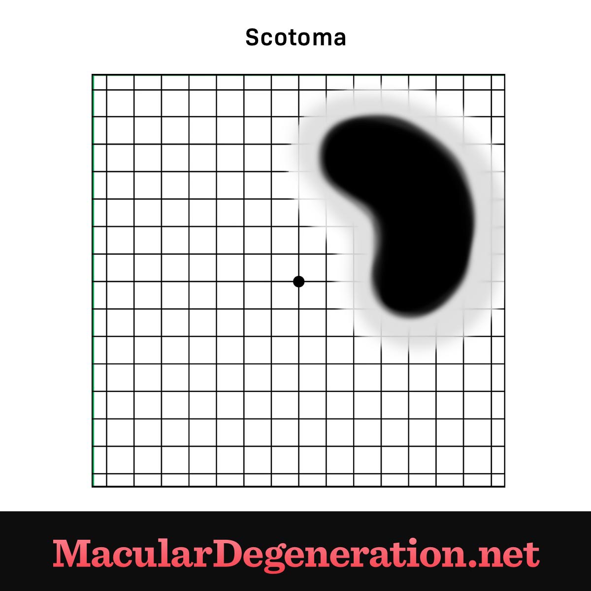
A scotoma is a blurry or blind spot in your visual field while the surrounding areas appear normal. A scotoma can appear in the center of your vision or along the periphery, and can be isolated, clustered, or spread out, and can also be present in just one eye or both.1
With macular degeneration, a scotoma most often appears in the central visual field, so it is called a central scotoma. That’s because in dry AMD, the light-sensing cells (called rods and cones) in center of the retina degenerate along with their supporting cells.2 In wet AMD or those with choroidal neovascularization (CNV) in myopic macular degeneration, there may be a leakage of fluid or bleeding underneath the retina.
How scotomas occur
The macula is the central area of the retina and is located in the back of your eyeball. The retina contains multiple layers of cells; one of these layers senses light and transmits visual signals to the brain through the optic nerve.
In dry age-related macular degeneration, light-sensing cells degenerate over time along with their supporting cells. Once enough cells in the macula degenerate, a blurry or blind spot can appear in the center of your vision, called a scotoma. In wet AMD or those with CNV in myopic macular degeneration, central scotomas occur from a sudden leakage of fluid or bleeding underneath the retina.
Dry AMD?
In early-stage dry AMD, the most common type of AMD, you may not notice any loss of vision.
In intermediate dry AMD, you may begin to notice a blurry or fuzzy pattern around the central part of your vision.3 As the disease progresses, that blurriness may turn into a central dark spot. At this stage, learning to shift your focus to your peripheral vision, utilizing brighter lights, and employing magnifying tools will help you read and perform other daily activities.
In advanced stage dry AMD, the scotoma can develop into an area of blurriness or darkness that becomes larger and more severe. At this point, it may become very difficult or impossible to read, drive, or discern faces.4
Wet AMD and myopic macular degeneration
Wet macular degeneration is another form of advanced or late macular degeneration and involves the growth of new abnormal leaky blood vessels underneath the central retina. The growth of abnormal leaky blood vessels underneath the central retina is called choroidal neovascularization (CNV), and can also occur in those with myopic macular degeneration.
Scotomas may develop in those with wet AMD or CNV in myopic macular degeneration. However, rather than the gradual blurring seen in dry AMD, individuals tend to develop central scotomas due to sudden leakage of fluid or bleeding underneath the retina.
Figure 1. Scotoma on an Amsler grid
Fortunately, prompt evaluation and treatment by an eye doctor can reduce the amount of fluid or blood, which can lead to a significant improvement in vision.
